Conquering Complexity: The ongoing revolution in oncology biomarker testing
 We are also in the midst of much more radical revolution, however. The molecular diagnostics landscape – and the very way we think about fighting cancer – is set to change dramatically in the coming decades.
In this update of our 2017 edition of this paper, we combine new data from Ipsos’ Global Molecular Diagnostics Monitors with our current therapy expertise and market insight to outline key developments in the oncology diagnostics landscape, and the resulting impact on cancer treatment.
We also offer a forward-looking perspective on how we believe this landscape will shift, and how today’s touchpoints may look very different in years to come.
Watch out for our upcoming podcast in which author, Pieter De Richter, talks us through the paper, offering his expert perspective on the future of oncology diagnostics. Details to follow.
Conquering Complexity: The Ongoing Revolution in Oncology Biomarker Testing
The Ipsos Global Oncology Centre of Expertise
1. On the cusp: from evolution to revolution The clinical management of cancer patients has undergone a remarkable evolution in the last 15 years, with the concept of personalised medicine now well-entrenched in the treatment paradigm across a wide range of tumour types, and it is becoming increasingly tumour-agnostic. Whereas treatment decisions used to rely on a combination of clinical observations, various macro-imaging techniques, and general histopathological findings, oncologists now have a range of molecular biomarker tests at their disposal to make a more informed drug choice. Notwithstanding the added complexity such testing brings1, this approach ultimately benefits physicians and patients alike: treatments which are likely to lead to better response rates and more prolonged responses can be selected based on molecular characteristics exhibited by the patient’s tumour. Cancer is a cunning adversary and fighting back requires an array of approaches that strike at the very heart of this foe: by targeting the mutations that drive and empower it, and by staying on top of the mutations that make it resistant to the weapons we employ. However, despite the advent of targeted therapies and the associated rise in the use of companion diagnostics, we are only just emerging from the initial exploratory stages of oncology biomarker testing. As will be argued in this paper, we are in the early phase of a much more radical revolution, and the molecular diagnostics landscape – and indeed the very way we think about fighting cancer – is set to change dramatically in the coming decades. It is crucial that any company entering this space prepare for this imminent upheaval and plan their launch strategy accordingly.
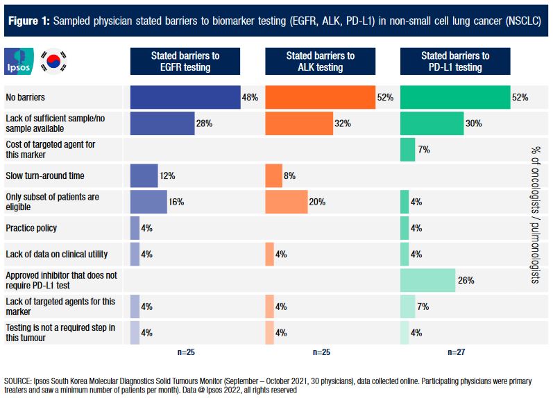
2. Phase change: from solids to liquids The central principle at the core of using molecular diagnostics to inform anti-cancer treatment decisions is that the patient’s tumour exhibits certain aberrant characteristics that predispose it to interventions at the protein and/or nucleic acid level. These characteristics can be either rare (e.g., ALK rearrangements, seen in 3-7% of NSCLC cancers2) or common (e.g., BRAF mutations, seen in ~50% of melanomas3), but the key here is that they are, by their very nature, not universal. In the majority of cases, these mutations, or abnormal expression patterns, are present only in the tumour cells: they are somatic mutations, not Conquering Complexity: The Ongoing Revolution in Oncology Biomarker Testing shared with the rest of the body’s cells, and absent from the germline (the body’s reproductive cells). This sets them apart from germline mutations, which are present in all of the patient’s cells. For the purposes of this discussion, we will focus on somatic tumour mutations, though most of the concepts in this paper also apply to inherited mutations. By definition, somatic mutations are not present in non-malignant cells in the patient’s body, and therefore it is no surprise that the traditional approach for tumour biomarker testing for patients with solid cancers has in the past relied on obtaining tissue samples from the actual tumour. This, unfortunately, comes with several drawbacks: initial biopsies used in diagnosis do not always contain enough viable tissue for testing (as highlighted in data deriving from Ipsos’ South Korea Molecular Diagnostics Solid Tumours Monitor - see figure 1), biopsy samples cannot be stored indefinitely without degrading, there is considerable intra-tumour genetic heterogeneity4, and re-biopsies mean the patient – often with a poor performance status – needs to undergo another invasive procedure. The latter point is particularly important when it comes to patients who have experienced a disease progression, which may be indicative of the tumour having acquired novel somatic mutations not present at the time the initial biopsy was taken.
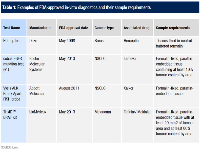
For some time, it seemed that this was to be accepted as an inherent limitation of solid tumour biomarker testing, and many tests were approved since the late 90s that relied on this principle of tissue-based testing. Below (Table 1) is a short, example list of several of the many FDA-approved biomarker tests for various solid cancer types, and their respective sample requirements:
It turned out, however, that it was possible to recruit one of cancer’s main weapons to help fight it. Solid tumours, like breast and lung cancer, begin as small groups of localised malignant cells in the primary organ in which they arise, but ultimately become much more dangerous to the host body by spreading to distant organs. Their relentless drive to replicate means that tumour cells often gain the ability to spread through the human body, in many cases (especially if not treated early enough) resulting in distant metastases. The way cancer metastasises is by shedding cells from the primary tumour, which enter the lymphatic system and/or the bloodstream5, and travel to distant sites where they eventually take hold and replicate. These so-called circulating tumour cells (CTCs) were first observed by Thomas R. Ashworth as early as 18696, and it was theorised that it should one day be possible to detect CTCs and use them for biomarker testing. Furthermore, the much more recent discovery of cell-free circulating tumour DNA For some time, it seemed that this was to be accepted as an inherent limitation of solid tumour biomarker testing, and many tests were approved since the late 90s that relied on this principle of tissue-based testing. Below (Table 1) is a short, example list of several of the many FDA-approved biomarker tests for various solid cancer types, and their respective sample requirements:
It turned out, however, that it was possible to recruit one of cancer’s main weapons to help fight it. Solid tumours, like breast and lung cancer, begin as small groups of localised malignant cells in the primary organ in which they arise, but ultimately become much more dangerous to the host body by spreading to distant organs. Their relentless drive to replicate means that tumour cells often gain the ability to spread through the human body, in many cases (especially if not treated early enough) resulting in distant metastases. The way cancer metastasises is by shedding cells from the primary tumour, which enter the lymphatic system and/or the bloodstream5, and travel to distant sites where they eventually take hold and replicate. These so-called circulating tumour cells (CTCs) were first observed by Thomas R. Ashworth as early as 18696, and it was theorised that it should one day be possible to detect CTCs and use them for biomarker testing. Furthermore, the much more recent discovery of cell-free circulating tumour DNA (ctDNA7) and RNA (ctRNA) opened up additional possibilities for detection of mutations in the blood, through so-called “liquid biopsies”.
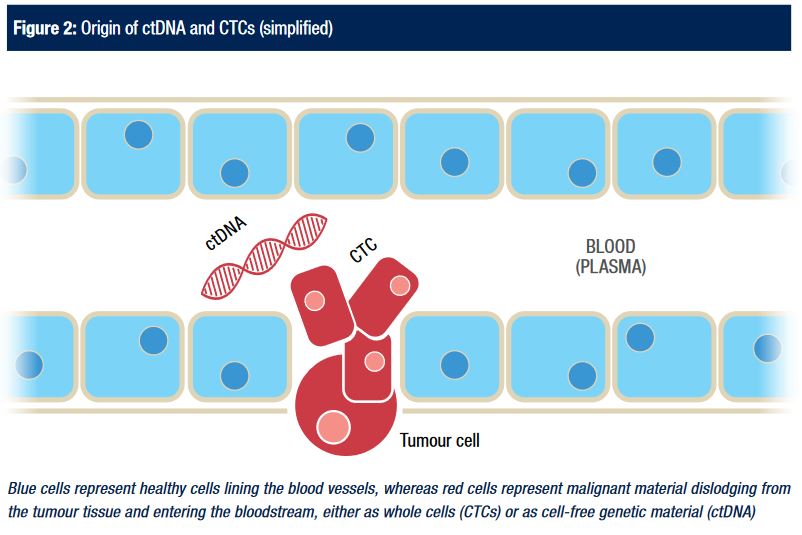
Blue cells represent healthy cells lining the blood vessels, whereas red cells represent malignant material dislodging from the tumour tissue and entering the bloodstream, either as whole cells (CTCs) or as cell-free genetic material (ctDNA). Before that could become a reality, several key challenges needed to be overcome: assays needed to be sensitive and specific enough to detect somatic mutations from very low concentrations of tumour DNA, against a background of non-malignant cells that far outnumber the cancerous cells/nucleic acid8. With the improvement in sensitivity of nucleic acid sequencing techniques and an associated drop in cost, this only became a practical possibility in the last few years. Indeed, the first half of the previous decade saw the beginnings of an increase in clinical trials that investigated testing CTCs and ctDNA for the purpose of selecting the most appropriate cancer treatment, and this culminated in the first FDA approval of a commercial liquid biopsy-based assay in June 2016: the Roche cobas EGFR Mutation Test v29. Prior to this date, the kit was already available and approved for detecting sensitising EGFR mutations in formalin-fixed paraffin-embedded (FFPE) tissue samples to identify patients eligible for treatment with erlotinib. The label extension allowed the kit to be used for identifying those mutations based on ctDNA. As the FDA approval at the time stated, “this new test may benefit patients who may be too ill or otherwise unable to provide a tumour specimen for EGFR testing.”
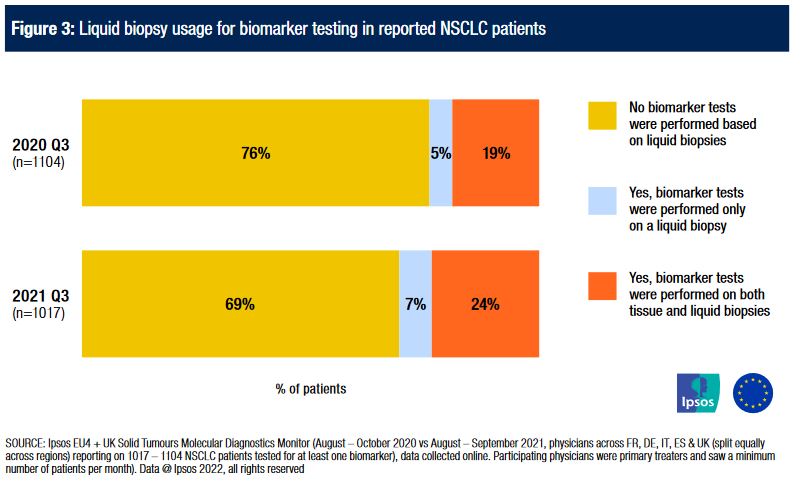
An important subsequent development was the further label extension of this test kit as a companion diagnostic for osimertinib three months later, based on the detection of T790M resistance mutations in blood or tissue samples11. This is significant, as the clinical and practical benefit of being able to test for (acquired) resistance mutations based on blood samples exceeds that of testing for (initial) sensitising mutations. As most testing for sensitising mutations is done at diagnosis, there usually is enough tissue available based on the initial biopsy (or resection) that was performed to confirm the cancer diagnosis and histology, limiting the need for blood-based tests (although there are theoretical advantages in turn-around time over tissue-based testing). On the other hand, testing for acquired resistance mutations should – by definition – be performed after treatment failure, which therefore requires a new sample to be collected in order to detect any newly acquired mutations. Furthermore, there may be a benefit to regularly conducting tests for resistance mutations such as T790M, to track the evolution of the genetic make-up of the tumour during its exposure to targeted therapies. With liquid biopsies removing many of the barriers relating to re-testing, and with an increasing number of third and fourth-generation targeted therapies designed to overcome resistance mutations available12, we are continuing to observe a steady increase in the use of liquid biopsies in the Ipsos EU4 + UK Molecular Diagnostics Solid Tumours data, as shown in Figure 3. It is important to note, however, that liquid biopsies are not yet replacing tissue biopsies: indeed, the majority of liquid biopsies are used for patients who have also had biomarker tests based on a solid biopsy. At this stage, they are mostly an additional tool, rather than a substitute for tissue biopsy-based testing.
Indeed, since 2015, a range of single-marker tests based on liquid biopsies have been made commercially available (some as non-FDA-approved lab-developed tests (LDTs)), covering a range of different markers and cancer types, as illustrated in Table 2:
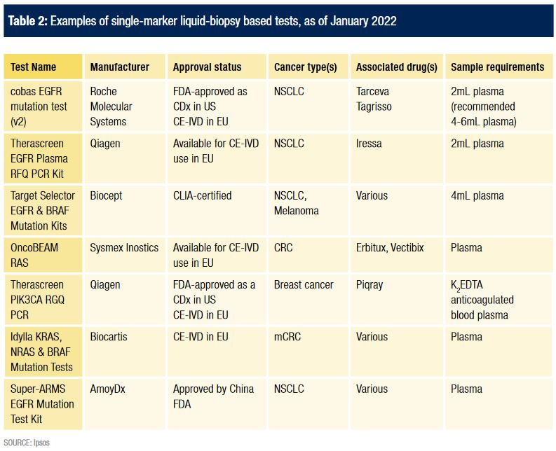
As innovative and promising as these tests are, despite the radically different sample requirements they still represent a fundamentally unchanged approach in terms of what markers are being tested: each of the tests listed above looks for mutations/changes in expression levels of single genes or proteins (though some commercial laboratories allow for multiple individual markers to be requested at the same time, provided there is enough liquid in the sample). Cancer, however, is a hugely complex disease (or set of diseases) and involves the interaction between many different such genes and proteins. Resistance – whether primary or acquired – is often the result of mutations in one, or several, of a long list of genes that would be very challenging to pick up through multiple single-marker test kits, much like looking for a needle in a genomic haystack.
3. Strength in numbers: from single-marker tests to whole genome panels (and everything in between)
The inherent limitations of single-marker tests are two-fold:
1. Genes do not act in isolation. The forces that drive the uncontrolled replication of cancer cells are far more complex than simple on/off switches. While a certain degree of success has been obtained by targeting specific, individual mutations in cancer cells, these approaches fail to address the fact that oncogenesis (the transformation of healthy cells into cancer cells) is a highly complex process that is usually the result of a combination of multiple mutations acting together, and that the kinases and other proteins that are encoded by proto-oncogenes and tumour suppressor genes form parts of intricate signal transduction cascades13 (signaling pathways within and between cells).
2. Not all cancers exhibit mutations commonly found in that cancer type. Clinical trials have understandably focused on mutations or amplification of genes that are relatively frequently observed in the population of interest. However, given the number of genes upstream and downstream of those common markers, just testing for those particular biomarkers would not identify any of the many potential rare abnormalities. By extension, certain mutations are very common in certain cancer types only (for example, KRAS mutations in colorectal carcinoma), but are rare in other cancers, and are therefore often bypassed in the testing process in favour of more common mutations in those cancers (for example, BRAF mutations in melanoma). The fact that they are less common in those cancers, however, does not mean they are non-existent: KRAS mutations can be found in approximately 0.7% of all melanomas.
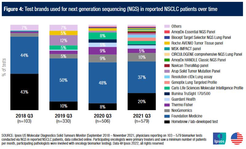
In the traditional testing paradigm that dominated much of the last decade, the only feasible way to overcome those limitations was to increase the number of single-marker tests performed on individual cancer tissues - rather than testing melanoma samples for just BRAF mutations, for example, separate BRAF, KRAS, NRAS and PIK3CA mutation tests could be performed to detect less common aberrations and to gain more insight into the specific molecular characteristics of that patient’s cancer. However, this approach presents several challenges: it requires more viable tumour tissue, it requires more time and effort, it significantly complicates the testing workflow and reagent/assay requirements, and multiplies the cost of testing every time an additional marker is added. Because of those limitations, using this approach beyond four or five different genes is usually not practically feasible. The rise of next generation sequencing (NGS) – driven by the continued drop in sequencing costs, down to as little as $0.005 per megabase in late 2021, from around $0.05 in early 201515 - has resulted in a way to approach this problem from a different angle. Rather than testing each gene with an individual assay, gene panels run on highly specialised equipment aim to sequence a large number of genes simultaneously. The resulting output then provides the physician with a mutational status for each of those many genes in one go, greatly increasing the available information regarding that patient’s tumour. Until relatively recently, the cost and complexity of NGS testing panels meant that they were mostly Conquering Complexity: The Ongoing Revolution in Oncology Biomarker Testing limited to somewhat experimental panels in large academic institutions. In the past decade, however, numerous diagnostics companies have launched and marketed commercial testing panels as integrated solutions: doctors are able to send samples for analysis to those companies’ dedicated laboratories, and will receive - often very detailed - reports on the genetic characteristics of the tumour, complete with treatment and/or clinical trial recommendations. What’s more, in addition to some of those panels now having approval as companion diagnostics for specific drugs in specific cancer types, many are now marketed as tumour-agnostic testing panels, meaning that they can be used for any of a wide range of cancers, since they cover such a large number of genes, many of which play a role in multiple tumour types. In fact, since 2017’s approval of pembrolizumab in mismatch repair-deficient (MMR-D) patients16 (regardless of cancer type), the FDA has now approved several other drugs for tumour-agnostic indications17,18, based on genetic abnormalities (in particular NTRK fusions) detected with NGS sequencing panels. The number of commercial pan-cancer testing panels has undergone an explosion in the last few years. This is reflected in data arising from Ipsos’ US Molecular Diagnostics Solid Tumours Monitor, which shows how the NGS testing landscape in the non-small cell lung cancer (NSCLC) market in the reported sample has expanded from just one main commercial provider in 2018 Q3 to a long list of panels, most with <5% market share each (see figure 4). Also included is the aggregate total of in-house test panels (panels typically developed by academic institutions for use by their practice only and not available as branded solutions).
4. The sum of the parts: liquid NGS panels The two major new developments described in the earlier section – clinically useful liquid biopsies and affordable, actionable next generation sequencing panels – arrived on the scene at around the same time. Several diagnostics companies sensed a real opportunity here and started working on products that sat squarely at the centre of where those two new technologies converged: NGS panels based on liquid biopsies. The theory behind these is elegant in its essence: if circulating tumour DNA (or ctRNA) can be detected in a cancer patient’s blood, and if tumour DNA can be used to conduct NGS testing panels, then a cancer patient’s blood should be a viable sample type for conducting such large multi-gene panels. Multiple technical hurdles had to be overcome before these liquid biopsy test panels could become a reality, however. The key challenge to Next Generation Sequencing of ctDNA is that the concentrations of mutated DNA fragments are typically so low (an order of magnitude lower than in cancer tissue samples) that the signal is obscured by the noise inherent in NGS machines. This meant that dramatic improvements in specificity were required to move beyond single marker testing towards sequencing complete exons in multiple gene targets19. Various diagnostics manufacturers have now overcome these challenges, and various such liquid biopsy testing panels are now available at commercial laboratories. Some examples are listed below:
- Guardant Health Guardant36020
- Guardant Health Guardant OMNI21
- Foundation Medicine FoundationACT22
- Illumina TruSight Oncology 500 ctDNA23
- Resolution Bioscience Resolution ctDx-Lung24
- NeoLAB Solid Tumor Liquid Biopsy25
As of late 2021, data from Ipsos’ US Molecular Diagnostics Solid Tumours Monitor showed that commercial liquid biopsy testing panels made up <10% of all commercial NGS testing panels (Fig. 5) in NSCLC (and other cancer types) in the reported sample. However, their use in the data has quadrupled since 2019, and continues to trend upwards. It is unclear when they may overtake solid biopsy-based NGS panels, but it is clear that they are shaping up to be a powerful additional tool in oncologists’ toolkit.

5. Looking ahead: the coming molecular intelligence revolution The advances discussed in the previous chapters are, as revolutionary as they may be, still fundamentally variations on the same basic approach: cancer cells or their genetic material (either tissue-bound or suspended in a liquid) are extracted, sent for analysis to a laboratory and analysed by a pathologist or related specialty; after which a report is generated and sent to the oncologist, who is then required to make a treatment decision. While immensely useful from a clinical perspective, this approach is far from perfect, due to a number of fundamental limitations:
i. Cancer cells are in a constant state of flux, by definition undergoing rapid division and often acquiring new mutations in the process26. Taking a snapshot at one point in time, or even at various points in the treatment journey, is going to miss a lot of the dynamic detail and identify resistance mutations with a significant amount of delay.
ii. Genes do not act in isolation. Even the largest pan-cancer testing panels are limited by the number of genes they cover, and even full exome sequencing services usually do not cover epigenetic alterations such as DNA methylation and histone modifications.
iii. Even the most well-trained pathologists or oncologists are limited by the amount of information they can process; there is already a risk of information overload with large NGS panels, and this issue increases exponentially with the number of genes/codons that are added to a panel: coming up with an appropriate treatment for a cancer based on the mutational and expression status of 1,000 different genes is infinitely more complex than doing so based on a handful of genes.
iv. Biomarker testing – whether single marker or multi-gene, whether tissue-based or liquid-based – is currently reactive: apart from hereditary (germline) panels to assess cancer risk, testing is conducted after a cancer diagnosis has been confirmed, and often after it has already metastasised. Detecting proto-oncogene or tumour suppressor gene mutations in the blood as soon as they arise could theoretically act as an early warning system prior to any symptoms presenting.
Addressing each of the barriers above will require a number of technological advances, but there are clear indications that the whole field of cancer biomarker testing is going to be radically different in the coming decades. Three major areas of research are likely to make a significant impact:
1. Labs on a chip, with remarkable sensitivities, which may ultimately be implanted in patients’ bodies/ bloodstreams, providing a means to continuously monitor biomarker status in real time. Taken one step further, these may be implanted prior to diagnosis, allowing cancer cells to be detected when they first arise.
2. Further improvements in the cost, speed and reliability of whole genome and whole exome sequencing. Following on from Next Generation Sequencing, interest in so-called “Third Generation Sequencing” is accelerating.
3. Perhaps most significantly, the inexorable rise of Artificial Intelligence (AI). In order to make sense of the enormous amount of data collected by whole genome/exome sequencing, especially when done so on a (semi-)continuous basis, human-based interpretation will necessarily need to be complemented with AI systems. NGS already utilises machine learning-enabled tools to ensure accurate read alignment, variant detection and variant annotation29, and machine learning/ deep learning will play an increasingly important role in interpretation and decision-making. Indeed, AI is already starting to prove itself in helping to recommend successful treatment combinations for specific molecular subtypes, as shown in a recent in vitro study in metastatic BRAF-mutated melanoma.
It is therefore not inconceivable that, at some point in the not-too-distant future, highly sensitive chips in individuals’ bodies will detect somatic mutations (or changes in gene expression patterns) as they arise, send this information in real time to powerful AI software, which will recommend a clinical course of action to be undertaken based on the wealth of molecular diagnostics information continuously transmitted through the chips’ sensors. This approach would represent personalised medicine taken to the extreme, and these systems would even continue to improve further through deep learning algorithms. The next – and perhaps ultimate - step in the more distant future would be pairing this ability with nanobots that permanently inhabit individuals’ bodies, dispensing targeted doses of highly specific drug cocktails, or physically destroying wayward cells, in an extremely localised manner in response to the information passed to them through the chips and AI agents. In this bold - but by no means unrealistic - view of the future, cancer would be destroyed before it has a chance to take hold, meaning it would effectively be cured. While this approach remains a largely theoretical future concept for now, the first small-scale human trials investigating the use of drug-delivering nanobots are already underway31. Indeed, nanobots are also currently being researched for the purposes of isolating and capturing circulating tumour cells32, which could theoretically be useful both for early detection and early treatment. In such a world, where nanobots and implanted chips collaborate to detect, isolate, capture, sequence and target malignant cells and their genetic material, constantly clearing our bodies of cells that exhibit mutations in proto-oncogenes or tumour suppressor genes, there would no longer be a need for biopsies, not even liquid biopsies. Biomarker testing would happen in real-time within our own bodies, and the involvement of reference laboratories, pathologists and even oncologists would essentially come to an end. Such a scenario is not yet around the corner, of course, and there are a great many obstacles to be overcome before we get there. Thanks to the advances of the last couple of decades, however, we are well on our way to conquering the complexity that makes curing cancer so difficult. Regardless of where the next decades take us, one thing is for certain: we are only in the early stages of an accelerating revolution in molecular diagnostics. In the five short years since the first version of this paper was written, the landscape has already changed very significantly. To assume that the same logistical touch points, stakeholders, market forces and dynamics that shape the oncology molecular diagnostics (MDx) market now are going to remain relevant in the coming decades would be very risky indeed.
About the Research
The Ipsos Global Molecular Diagnostics Solid Tumours Monitor is a multi-stakeholder, physician-reported syndicated patient and laboratory record database, capturing perceptions towards and usage of molecular diagnostics tests in solid cancer types. Participating drug-treating physicians are screened for specialty, level of seniority and number of drug-treated cancer patients seen per study wave and must be the primary decision-maker for their patients. In addition, participating pathologists must be involved with preparing samples, ordering cancer-related molecular diagnostics tests and/or performing/interpreting cancer-related molecular diagnostics testing in solid cancers, and must be aware of the methodology and/or brand used for cancer-related molecular diagnostics tests. Each wave, participants complete a perceptual usage and attitudes questionnaire, before providing de-identified information on a predefined quota of oncology patients seen in consultation / laboratory samples handled in practice, retrospectively (across a pre-defined list of solid tumour types). Data used in this article were collected online in US, France, Germany, Italy, Spain and the UK, and collected both online and via pen & paper in South Korea. Sample sizes are provided alongside the relevant charts.

![[WEBINAR] Cracking the Nutritional Supplement Market](/sites/default/files/styles/list_item_image/public/ct/event/2026-05/thumbnail-cracking-nutritional-supplement.png?itok=Cts0kD4T)
![[WEBINAR] Dealmaking 2026: What’s Changing, What’s Not, and How to Position for the Next Wave](/sites/default/files/styles/list_item_image/public/ct/event/2026-03/deal.png?itok=NtU1lGCc)